Why Use Microscopes?

Using microscopes during hair transplant surgery has allowed transplant results to evolve from the unnatural pluggy doll’s hair appearance to the more refined and natural results created by follicular unit transplantation.
The unnatural appearance of the early transplants was due to the larger grafts that contained several follicular units leaving a stalky and artificial appearance. Hair transplant surgeons then transitioned to micrografts, which were smaller than the larger 4 to 5 mm plugs but still did not deliver the refinement physicians and patients desired. This goal was finally achieved when a pioneering physician, Dr. Limmer, championed using microscopes for graft preparation. Microscope-aided graft dissection helped to make the large and unnatural-looking plugs and micrografts become ancient history.
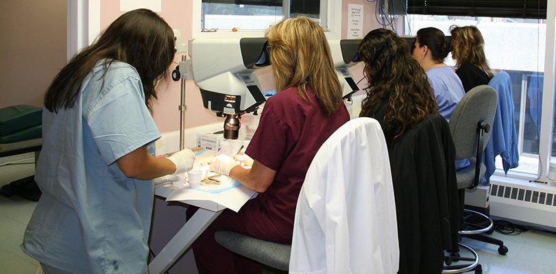
Our clinic uses high-powered stereomicroscopes because they provide unparalleled views of the follicles. Our team of technicians is better able to see the natural follicular groupings under the 3D high-powered magnification, which allows for precise dissection of the grafts as they are separated into their individual units from the graft harvest strip from a FUT procedure. These special microscopes also give the technicians better visualization of lighter-colored and grey hairs, thereby minimizing damage to these otherwise difficult-to-see follicles. This augmented visualization of the follicle also minimizes damage to the surrounding delicate support structures necessary for the graft to survive and grow. Our clinic also inspects all FUE grafts to assess for quality and likelihood of survival before being implanted. There are higher yields of usable grafts from the harvested strip with microscope-aided dissection because follicles are not wasted by being transected and cut into fragments that may not be viable for transplantation. Our skilled team of technicians consistently provides a high yield of high-quality, usable grafts.
The use of microscopes during hair transplant surgeries has improved graft quality and graft survival. This is crucial because donor follicles are of limited supply, and the use of stereomicroscopes optimizes graft survival and minimizes tissue loss and waste. The preservation of follicles during graft preparation allows us to maximize the use of a limited donor supply. Using stereomicroscopes for graft preparation and evaluation has proven invaluable when the goal is to have good-density hair transplant results that are natural looking and undetectable.


